Copy link
Anatomy and Physiology of Neuromuscular Transmission
Last updated: 06/04/2025
Key Points
- The neuromuscular junction is responsible for the chemical transmission of electrical impulses from nerves to muscles to generate a muscle contraction.
- The three main elements of the neuromuscular junction are the presynaptic motor terminal, the synaptic cleft, and the postsynaptic muscle endplate.
- Acetylcholine (ACh) is the major neurotransmitter that is released by the motor neurons.
- Upon binding of ACh to nicotinic ACh receptors on the postsynaptic membrane, receptor-associated sodium and potassium channels open, depolarizing the membrane via sodium influx and potassium efflux. If the depolarization reaches a threshold, adjacent voltage-gated sodium channels open, creating a muscle action potential that propagates to the sarcolemma and facilitates muscle contraction.
Anatomy and Physiology of Neuromuscular Transmission
- The neuromuscular junction is responsible for the chemical transmission of electrical impulses from nerves to muscles to generate a muscle contraction.
- The three main elements of the neuromuscular junction are (Figure 1):
- Presynaptic motor terminal
- Synaptic cleft
- Postsynaptic muscle endplate
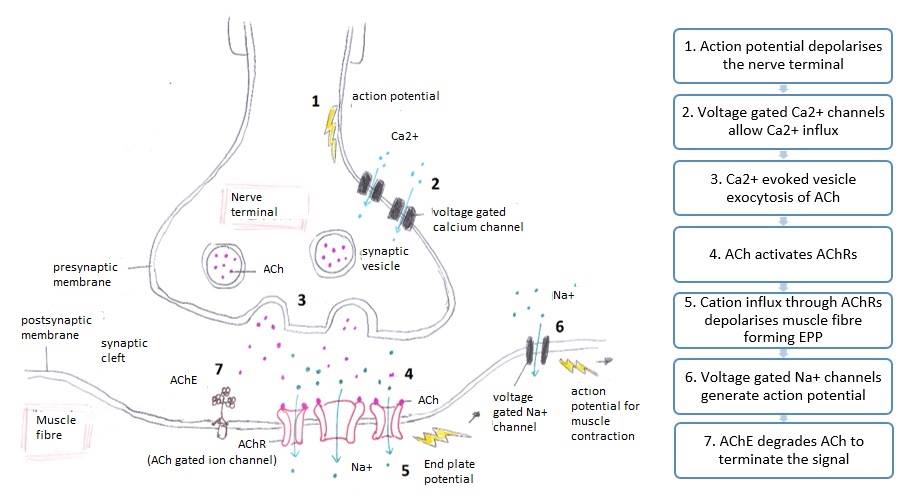
Figure 1. Neuromuscular junction anatomy and signaling. Source: Wikimedia Commons. 2014. Accessed May 23, 2025. Link
Presynaptic Motor Terminal, Motor Nerve Depolarization, and ACh Release1,2
- The presynaptic motor terminal is the end terminal axon of the motor nerve that communicates with the muscle endplate.
- It contains synaptic vesicles filled with ACh, voltage-gated calcium channels, and proteins for active vesicle docking and release.
- When the presynaptic motor terminal is depolarized via an action potential, voltage-gated calcium channels open in response, resulting in calcium influx into the cell (steps 1 and 2 of Figure 1).
- Calcium influx results in exocytosis of ACh-containing vesicles from the presynaptic membrane into the synaptic cleft (step 3 of Figure 1).
Synaptic Cleft1,2
- The synaptic cleft is a narrow extracellular space (about 50 nm) through which neurotransmitters, such as ACh, travel to reach their receptors on recipient nerves or muscle endplates.
- The synaptic cleft of the neuromuscular junction contains acetylcholinesterase, which degrades ACh that was released from the presynaptic terminal (see step 7 of Figure 1).
Postsynaptic Muscle Endplate1,2
- The postsynaptic muscle endplate is the membrane of the recipient muscle fiber that is depolarized by ACh release from the presynaptic nerve.
- The postsynaptic membrane has multiple folds located opposite the presynaptic nerve terminal.
- Upon binding of ACh to nicotinic ACh receptors on the postsynaptic membrane, receptor-associated sodium and potassium channels open, depolarizing the membrane via sodium influx and potassium efflux. If the depolarization reaches a threshold, adjacent voltage-gated sodium channels open, creating a muscle action potential that propagates to the sarcolemma and facilitates muscle contraction (see steps 4-6 of Figure 1).
Muscle Excitation and Contraction3
- The electrical depolarization from the neuromuscular junction travels to the sarcolemma and into the T-tubules, which releases calcium from the dihydropyridine receptors.
- The calcium from dihydropyridine receptors then binds to ryanodine receptors, causing further calcium ion release from ryanodine receptors in the sarcoplasmic reticulum (Figure 2).
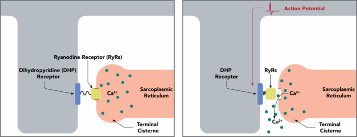
Figure 2. Coupling of the muscle action to Ca++ release from the sarcoplasmic reticulum: The dihydropyridine receptor and ryanodine Receptor. Source: Daniel Wash and Alan Sved, Wikimedia Commons. 2019. Accessed May 23, 2025. Link
- Calcium release from the sarcoplasmic reticulum binds to troponin C, which facilitates the movement of tropomyosin to expose actin binding sites for myosin heads to attach. This allows for myosin to bind to actin, facilitating muscle contraction.
- After attachment of the myosin head to actin, adenosine diphosphate and inorganic phosphate are released, causing a power stroke.
- Attachment of adenosine triphosphate (ATP) will cause the release of myosin from actin. ATP hydrolysis causes the myosin head to be in the “cocked” position to re-attach to actin (see Figure 3).
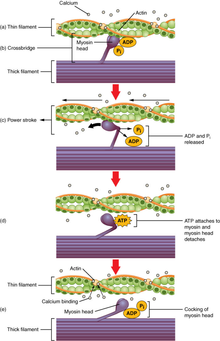
Figure 3. Skeletal muscle contraction. Source: OpenStax, Wikimedia Commons. 2016. Accessed May 23, 2025. Link
A general overview of neuromuscular depolarization leading to muscle contraction is illustrated in Figure 4.
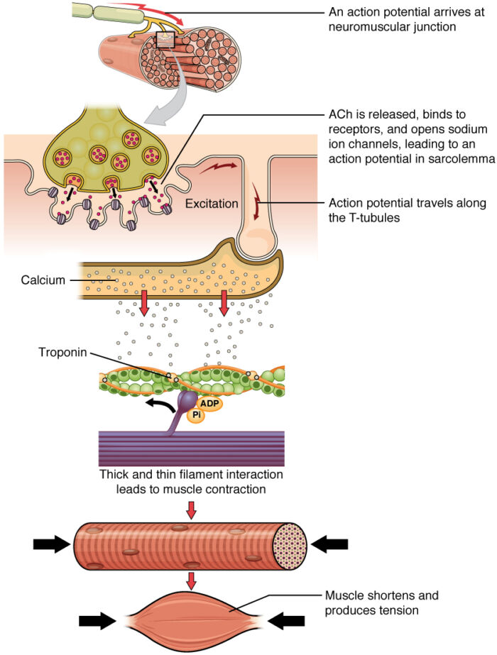
Figure 4. Generation of muscle contraction. Source: OpenStax, Wikimedia Commons. 2016. Accessed May 23, 2025. Link
ACh Synthesis and Breakdown4
- Ach is synthesized in the presynaptic terminal when choline is acetylated by choline acetyltransferase. ACh is then packaged into synaptic vesicles for exocytosis to the synaptic cleft.
- It binds to nicotinic ACh receptors on the motor endplate.
- Ach is degraded in the synaptic cleft by the enzyme acetylcholinesterase into acetate and choline. Choline is recycled back to the neuron.
Nicotinic and Muscarinic Modulation5
- The action potential triggering voltage-gated calcium channel and ACh exocytosis is regulated by presynaptic nicotinic and muscarinic receptors.
- Nicotinic receptors increase further ACh release via positive feedback
- Muscarinic receptors (specifically M2 receptors) inhibit further ACh release via negative feedback.
The Nicotinic ACh Receptor4,6,7,8
- The nicotinic ACh receptor is a ligand-gated ion channel that is composed of five subunits.
- The subunits can be identical (homomeric) or different (heteromeric), with over 17 different subtypes being known so far (α1–10, β1–4, γ, δ, ε) (Figure 5).
- The most common combination in the brain is α4β2 subtype.
- The α7 homomeric subtype, a calcium modulator, is prevalent in the hippocampus and cortex.
- The subunit combination determines the physiological and pharmacological traits of the receptors, including pharmacokinetics, agonist binding affinity, and ion selectivity.
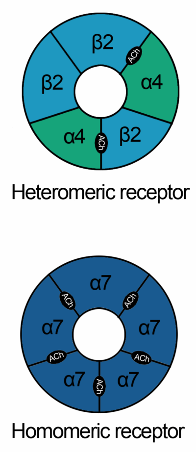
Figure 5. Heteromeric and homomeric nicotinic ACh receptors. Source: Wikimedia Commons. 2009. Accessed June 1, 2025. Link
- There are both presynaptic and postsynaptic nicotinic ACh receptors.
- Presynaptic nicotinic ACh receptors are distributed throughout the central nervous system and modulate neurotransmitter release (GABA, dopamine, glutamate, serotonin). This plays a crucial role in synaptic plasticity, attention, and reward pathways, influencing cognitive function.
- Postsynaptic nicotinic ACh receptors cause depolarization and excitation of action potentials in the neuromuscular junction, as well as playing a minor role in neuronal excitability in the central nervous system.
- Upregulation of nicotinic ACh receptor occurs with chronic nicotine exposure (e.g., cigarette smoking), especially in the α4β2 subtype.
- The upregulation leads to increased receptor density and alters function, which forms the basis behind nicotine dependence, addiction, and withdrawal.
- Nicotinic ACh receptors are also expressed in non-neuronal tissues such as epithelial cells and immune cells, where they play a role in inflammation and cell signaling.
References
- Davis LA, Fogarty MJ, Brown A, Sieck GC. Structure and function of the mammalian neuromuscular junction. Compr Physiol. 2022;12(4):3731-66. PubMed
- Ruff RL. Neurophysiology of the neuromuscular junction: overview. Ann N Y Acad Sci. 2003; 998:1-10. PubMed
- Gash MC, Kandle PF, Murray IV, et al. Physiology, muscle contraction. Updated 2023. In: StatPearls. Treasure Island (FL): StatPearls Publishing; 2025. PubMed
- Fagerlund MJ, Eriksson LI. Current concepts in neuromuscular transmission. Br J Anaesth. 2009;103(1):108-114. PubMed
- Tomàs J, Santafé MM, Garcia N, et al. Presynaptic membrane receptors in acetylcholine release modulation in the neuromuscular synapse. J Neurosci Res. 2014;92(5):543-54. PubMed
- Kalamida D, Poulas K, Avramopoulou V, et al. Muscle and neuronal nicotinic acetylcholine receptors. Structure, function, and pathogenicity. FEBS J. 2007;274(15):3799-3845. PubMed
- Dani JA, Bertrand D. Nicotinic acetylcholine receptors and nicotinic cholinergic mechanisms of the central nervous system. Annu Rev Pharmacol Toxicol. 2007;47:699-729. PubMed
- Kellar KJ, Dávila-García MI, Xiao Y. Pharmacology of neuronal nicotinic acetylcholine receptors: effects of acute and chronic nicotine. Nicotine Tob Res. 1999;1 Suppl 2:S117-S140. PubMed
Other References
- Neuroscientifically Challenged. Neuromuscular junction. Created 2016. Accessed June 4, 2025. Link
- Macias O, Cha YM. OA summary on Neuromuscular Blockade: Basics. Created April 26, 2023. Accessed June 4, 2025. Link
- Heng RM, Olbrecht VA. OA summary on Nondepolarizing Neuromuscular Blocking Agents. Created November 10, 2023. Accessed June 4, 2025. Link
Copyright Information

This work is licensed under a Creative Commons Attribution-NonCommercial-NoDerivatives 4.0 International License.