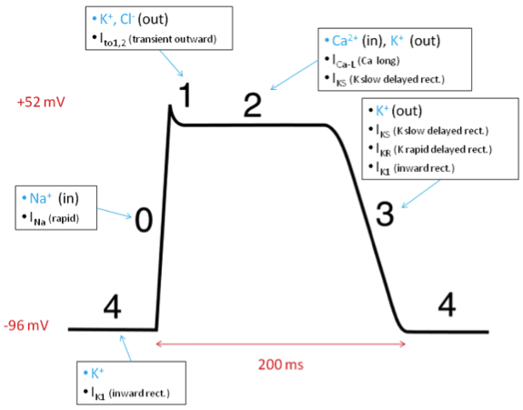
For skeletal muscle, Na+ mediated depolarization (Phase 1) is quickly followed by repolarization (Phase 3). Repolarization is mediated by closure of the Na+ channels responsible for depolarization, and opening of voltage-gated K+ rectifier channels, which allows for potassium efflux. These K+ channels will remain open and hyperpolarize the cell membrane is restored to its resting state (-90 mV).
Cardiac myocytes are more complicated. Depolarization is initially followed by a “plateau phase” (Phase 2) where the membrane potential remains positive for 0.2-0.3 seconds. During this phase, Ca2+ influx through L-type Ca2+ channels balances K+ efflux through slow delayed rectifier K+ channels. Eventually, the Ca2+ channels close while the K+ permeability increases as rapid rectifier K+ channels open, allowing for rapid repolarization (Phase 3). K+ channels remain open until the resting membrane potential is restored.

Copyright Information

This work is licensed under a Creative Commons Attribution-NonCommercial-NoDerivatives 4.0 International License.