Key Points
- The lateral surface of a 5-week-old embryo contains pairs of narrow masses called branchial (pharyngeal) arches that will develop into upper airway structures.
- The lower airway structures begin as an outpouching of the primitive foregut and continue to develop into young adulthood.
Upper Airway Development
The lateral surface of a 5-week-old 4.0mm embryo contains five or six pairs of narrow masses called branchial or pharyngeal arches. Each arch contains ectoderm, the precursors of the epithelial structures (e.g., skin), and mesoderm, the precursors of mesothelial structures (e.g., muscle, bone). The structures of each branchial arch receive motor or sensory innervation from an adjacent cranial nerve and retain this original embryonic innervation during migration.
The first branchial arch develops into the mandible, maxilla, muscles of mastication, bones of the middle ear, and the muscles between the ear and mandible. Motor and sensory innervation are supplied by the trigeminal nerve (cranial nerve (CN) V).
The second branchial arch forms muscular structures innervated by the facial nerve (CN VII), including muscles of the face and inner ear, the styloid process, and lesser cornu of the hyoid bone.
The third branchial arch develops into the body and greater cornu of the hyoid bone and the stylopharyngeus muscles, which elevates the palate during swallowing and is innervated by the glossopharyngeal nerve (CN IX).
The fourth through sixth branchial arches contribute to the formation of the thyroid, cricoid, arytenoid, corniculate, and cuneiform laryngeal cartilages as well as the muscles that form the pharynx, larynx, and upper half of the esophagus. The innervation to these structures are from the vagus (CN X) and accessory (CN XI) nerves.1

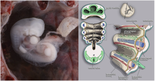
Figure 1a. Embryo at 4 to 5 weeks showing pharyngeal arches Source: lunar caustic. CC BY 2.0 (left), 1b. Pharyngeal Arch Human. Source: Loki austanfell, Wikimedia. CC BY SA 3.0 (right).
Lower Airway Development
Lower airway development begins as the embryonic stage, at 4-7 weeks gestation as a ventral outpouching of the primitive foregut, which is the primitive larynx and trachea. A pair of tubes, the lung buds, form at the posterior end of this outpouching, which will ultimately form the lungs. Esophagotracheal septum or ridge forms, dividing the primitive foregut into the esophagus and trachea. During the pseudoglandular stage, the lung buds grow, branch and begin to proliferate. Throughout the canalicular stage, the respiratory bronchioles develop, budding off from the terminal bronchioles. Organogenesis is complete with the formation of the pleura.2
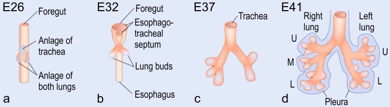
Figure 2. Early human lung development. Source: Cell Tissue Res. 2017; 367(3): 427–444. CC BY 4.0.
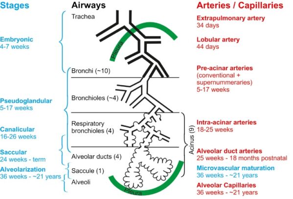
Figure 3: Overview of lower airway development Source: Cell Tissue Res. 2017; 367(3): 427–444. CC BY 4.0.
During the saccular stage, the terminal airways grow in length and width and form a cluster of larger airspaces, or sacculi. During this stage, there is a decrease in interstitial tissue and a thinning of the walls. Finally, true alveoli form at around 36-weeks gestation, initiating the alveolar stage of lung development, which continues into young adulthood.3
References
- Isserman RS, Litman RS. Developmental anatomy of the airway. In Narasimhan J and Fiadjoe JE, Eds. Management of the Difficult Pediatric Airway. Cambridge UK; Cambridge University Press; 2020: 1-7
- Schittny JC. Development of the lung. Cell Tissue Res. 2017; 367(3):427-444. PubMed
- Burri, PH. Structural Aspects of Postnatal Lung Development – Alveolar Formation and Growth. Biol Neonate. 2006; 89:313-322. PubMed
Copyright Information

This work is licensed under a Creative Commons Attribution-NonCommercial-NoDerivatives 4.0 International License.