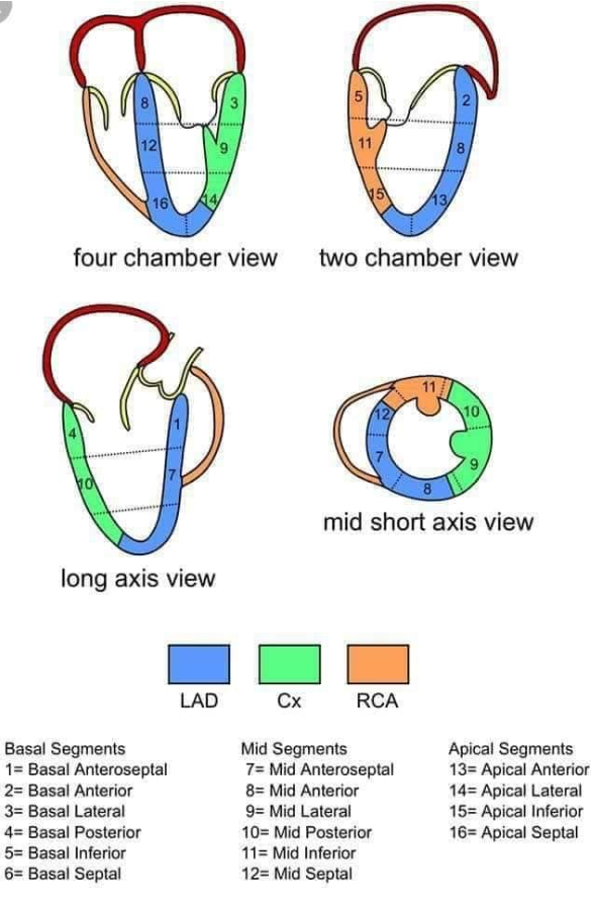
Mid Esophageal 4 Chamber View: In this view, the lateral wall of LV as well as the septal wall are visualized. The lateral wall is supplied by the left circumflex artery (LCX) and the septal wall is supplied by perforating branches of the left anterior descending artery (LAD) and the posterior descending artery (PDA). You can also see the A2 and P2 segments of the mitral valve with A2 closest to the LVOT. Pulling the probe out from this position will yield the 5 chamber view which is notable for the whole LVOT and aortic valve.
Mid Esophageal 2 Chamber View: This is obtained from the 4 chamber view by increasing the omniplane angle to approximately 90o. This vertical slice of the LV shows the anterior wall on the right of the image and the inferior wall on the left. The A1, A2, and P3 segments of the MV can be seen in this view with A1 closest to the anterior LV wall (image right).
Mid Esophageal Commissural View: This is between the ME4C and the ME2C view with the omniplane angle between 45-60. You can see the Anterolateral (right) and Posteriomedial (left) papillary muscles of the mitral valve in this view and can assess for papillary rupture. You can also see P1 (right) P3 (left) and M2 (“trap door” in the middle) scallop leaflets of the mitral valve in this view.
Mid Esophageal Long Axis View: The MELAX is obtained by turning the omniplane to around 130. The Anterioseptal wall (image right) and inferolateral walls of the LV can be assessed in this view. The LVOT is clearly seen and the aortic valve can be assessed for the presence of aortic insufficiency in this view. The A2 leaflet is closest to the LVOT and the P2 leaflet of the mitral valve is seen on the left of the image. By turning the probe slightly to the patient’s right, the RV can be seen and the septal wall can be assessed.
Transgastric Short Axis: The LV will be viewed as a donut in this view with the inferior wall closest to the transduce (top of image). The lateral wall is on the right and the septal wall is on the left. The anterior wall is on the bottom of the screen. The papillary muscles can be seen in this view. This is a useful view because all coronary arteries can be assessed in one view: the LAD supplies the anterior wall and anterior 2/3rds of the septum, the LCX supplies the lateral wall, and the PDA supplies the inferior wall. Aggressive antiflexion can show the fish mouth of the mitral valve. Sliding the probe slightly deeper allows for assessment of the apex.
Deep Transgastric Long Axis: This view is at an omniplane of o and images the LV through the apex and allows for assessment of the aortic valve which is the Left of the 2 valves seen with the mitral being the right most. Here, continuous wave Doppler can assess the AV for either regurgitation or stenosis.
History: 51%; 2018
Author: Dan Smith, MD