The peripheral nervous system is divided into somatic and autonomic components. The somatic nervous system includes the sensory and motor nerves that innervate the limbs and body wall. Sensory nerve fibers in the peripheral nerves are the peripheral axonal process of neurons in the dorsal root ganglion. The motor axons are the processes of anterior horn cells of the spinal cord.
Nonmyelinated nerves, such as autonomic postganglionic efferent and nociceptive afferent C fibers, contain many axons encased in a single Schwann cell sheath.
All large motor and sensory fibers are enclosed in many layers of myelin, which consists of the plasma membranes of specialized Schwann cells that wrap themselves around the axon during axonal outgrowth.
Myelin greatly increases the speed of nerve conduction by insulating the axolemma from the surrounding conducting salt medium and forcing the “action current” generated by an impulse to flow through the axoplasm to the nodes of Ranvier, which are periodic interruptions in the myelin sheath where the active impulse is regenerated . The Na+ channels that serve generation and propagation of impulses are highly concentrated at the nodes of Ranvier of myelinated fibers[7] but are distributed all along the axon of nonmyelinated fibers.
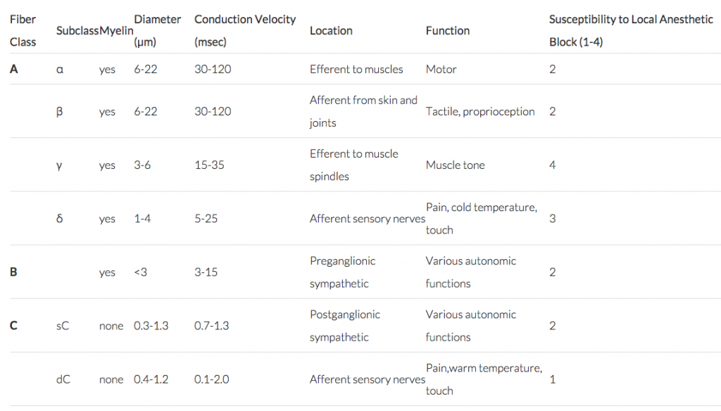
A classification of peripheral nerves according to fiber size and physiologic properties (see image).
A typical peripheral nerve consists of several axon bundles, or fascicles. Each axon has its own connective tissue covering, the endoneurium. Each fascicle of many axons is encased by a second connective tissue layer, the epithelial-like perineurium, and the entire nerve is wrapped in a loose outer sheath called the epineurium (Fig. 30-4). To reach the nerve axon, a local anesthetic molecule must traverse four or five layers of connective tissue or lipid membranous barriers, or both.

Copyright Information

This work is licensed under a Creative Commons Attribution-NonCommercial-NoDerivatives 4.0 International License.